
PIC Dental Technology
PiC camera
- One minute capture
- Patient move allowed
- Photogrammetry technology
PiC transfer
- All implant platforms
- Sterilizable 1.000 cycles
- Screw retained coded transfer
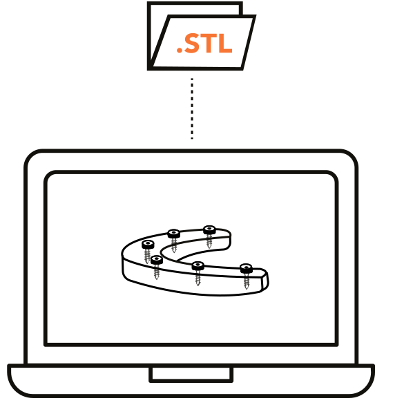
PiC file
- Open STL file
- CAD/CAM system integration
- Unalterable interrelated positions
PiC pro
- CAD/CAM enhancer
- Efficient Full Arch workflow
- Proprietary prostheses protocols
Implants capture
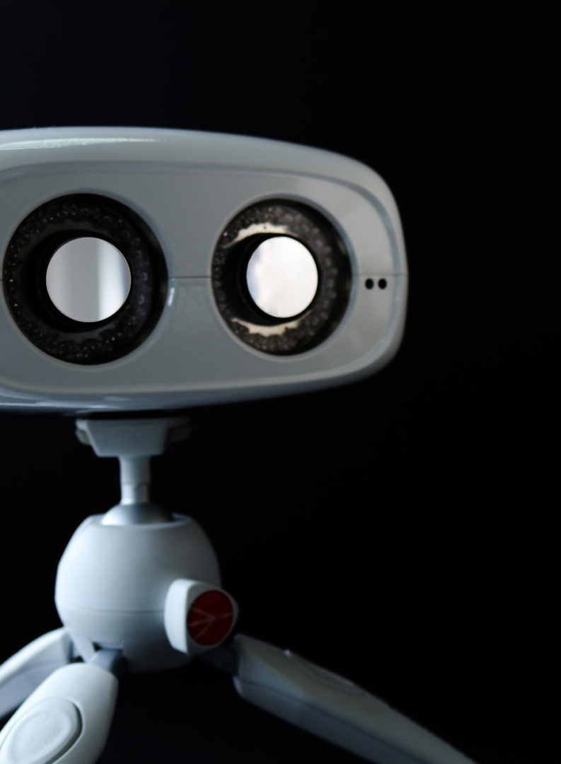
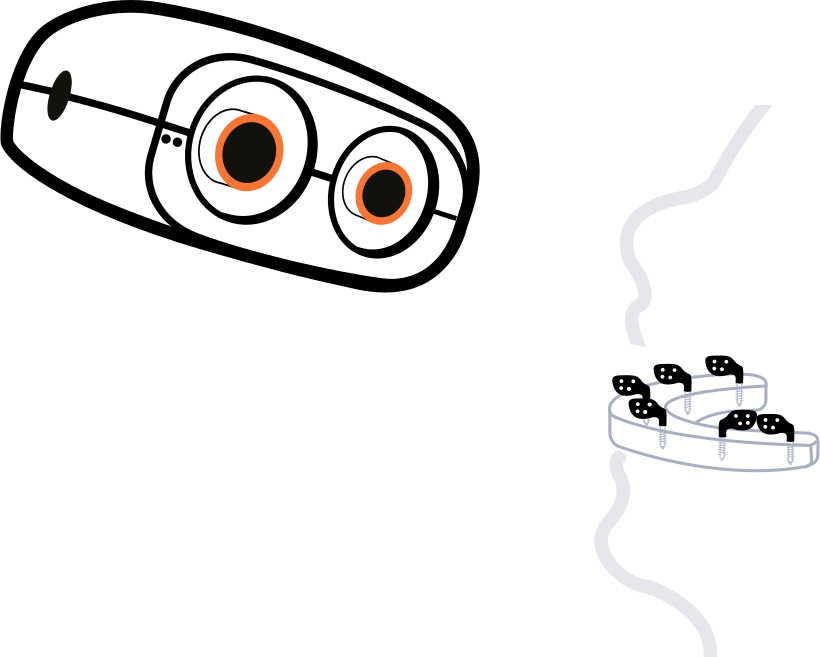
Screw the PiC transfers onto the patient’s implants and bring the PiC camera closer to obtain the implants positions in less than a minute.
PiC transfers are precisely placed, prior to capturing the measurements, on top of the implants or multi-unit abutments immediately after surgery or post healing as a step in the restorative phase. PIC transfers have a unique and customized pattern consisting of pre-measured marker dots. PiC dental system use photogrammetry technology to identify the vectors of each implant on an arch creating the most precise measurements which are interrelated. This allows for implant impressions to be captured among all implants digitally with up to 10 micron accuracy cross arch.
PiC camera and Software uses unique logarithms capable of full arch measurements in less than 1 minute. Patient information including case notes, implant positions along with system, size, healing cap and tooth position are entered into the system before measurements are taken. Once the procedure begins a viewer window opens on the computer screen to assist the user in capturing the precision measurements. PiC dental system is built to detect and alert if a PiC transfer shifts or was incorrectly selected ensuring a confirmed scan at completion.

Soft tissue registration
The next step is the acquisition of the contours of the soft tissues and the adjacent teeth via conventional impression or an intraoral scanner.
The normal procedure is to scan the soft tissues with the healing caps, so they will also be introduced in the PiC file to correlate the soft tissue data with the positions of the implants via Best Fit registration. The use of an intraoral scanner allows you to enter a digital workflow by scanning all the patient data needed for all implant restoration treatments. Sometimes, as in the case of the fully edentulous lower jaw, it is difficult to obtain a good impression of the soft tissues with an intraoral scanner due to moving parts such as the floor of the mouth or the vestibular areas near the lips, so that you take an alginate (keeping static the movable parts) and scan later this alginate with your iOS. This procedure is faster and more comfortable for the patient while the tuberosities and the alveolar ridge are better registered.
When taking a conventional impression to acquire the soft tissues, impression transfers are not needed, only healing caps (or Scan Bodies) must be visible over the gingiva in order to be correlated with the PiC file. If done on day of surgery then additional soft tissue impression will be necessary just after the suturing to detect changes occurring from healed tissue prior to delivering the final prostheses. Regardless of detection process, conventional impression or intraOral scanner, soft tissue is then superimposed with the PiC file to provide the world’s most accurate and finest restoration.
Precise digital model
The PiC file with the implants positions interrelated and the soft tissue registration are aligned in a dental CAD softwareScrew the PiC transfers onto the patient’s implants and bring the PiC camera closer to obtain the implants positions in less than a minute.
The last step of the PiC dental system is the creation of a digital master model. The introduction of digital workflows where your patient is scanned and displayed virtually on a screen changes the need to have a perfect master model. The prostheses are manufactured by milling machines while the models are manufactured in 3D printers, so there may be an accuracy discrepancy between the model and the prosthesis.
The virtual model is now more accurate than the physical model due to the addition of manufacturing errors through 3D printing or milling. Although milling machines used in laboratories and milling centers are more accurate than 3D printers, they always add a small manufacturing error, and although today the accuracy of 3D printers is improving, some error will always be added to the design of the prosthesis with respect to the scanned starting data.
The PiC Master model is manufactured combining the 5-axis milling machine and the 3D printer to have all the advantages of both manufacturing technologies.